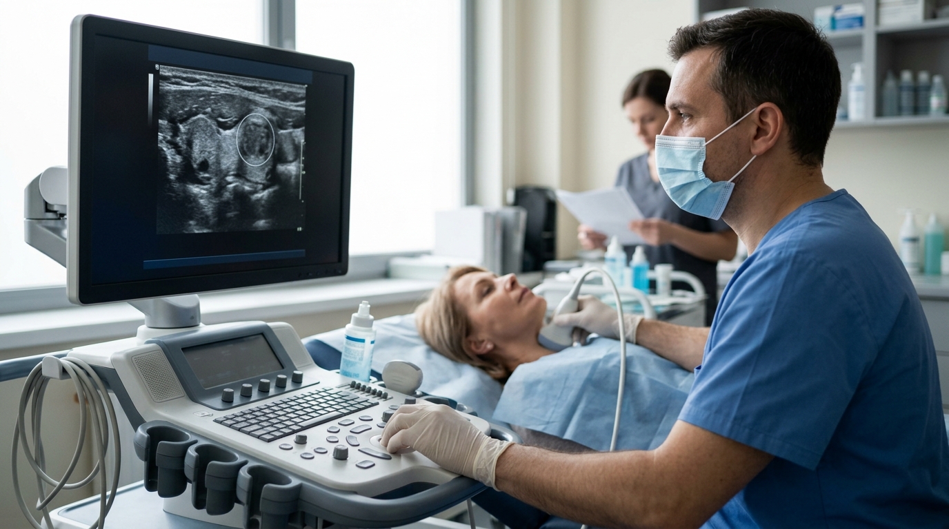
Your report describes a nodule by its size in millimeters and its ultrasound features, and that description is what decides whether anything follows.
A thyroid nodule is an area of tissue inside the thyroid gland that looks different on ultrasound from the tissue around it. Finding one is common: nodules are seen in as many as 68% of high-resolution ultrasound scans (American Journal of Roentgenology (ACR TI-RADS Committee), undated), and general medical consensus holds that more than 90% of thyroid nodules found in adults are benign (Cleveland Clinic, undated).
A nodule on a scan is a description of what the sound waves showed, not a diagnosis. What happens next depends on the size in millimeters or centimeters, the specific features the radiologist listed, and the recommendation written at the end of your own report — bring that report, not your memory of it, to your next appointment.
What This Finding Is
On ultrasound, the thyroid is a butterfly-shaped gland sitting low in the front of your neck, on either side of the windpipe. Normal thyroid tissue looks fairly uniform. A nodule shows up as a distinct patch within it — rounder or darker or brighter than its surroundings, sometimes filled with fluid, sometimes solid, sometimes a mix of both.
The radiologist measures it in three dimensions (reported in millimeters or centimeters) and describes its composition, how bright or dark it is, its shape, its edges, and whether there are tiny bright specks called calcifications. Those descriptions are what drive the recommendation, not the word "nodule" by itself.
As a general pattern, major health organizations report that most nodules are unrelated to cancer: more than 90% of thyroid nodules detected in adults are benign (Cleveland Clinic, undated), and thyroid nodules represent thyroid cancer in roughly 4.0% to 6.5% of cases (Cleveland Clinic, undated). Older primary-care literature puts malignancy at about 5% of all palpable nodules (American Academy of Family Physicians, 2003). Those are population patterns, not a prediction about your nodule.
Why This Is Sometimes Found
Common and mostly benign explanations first:
- A colloid nodule — an overgrowth of otherwise normal thyroid tissue. Major health organizations describe this as the most common type of thyroid nodule (Cleveland Clinic, undated).
- A cyst or partly fluid-filled nodule. Cystic nodules carry a low risk of malignancy and are generally either monitored or biopsied if larger than 2 cm (Cleveland Clinic, undated).
- Several nodules at once, often picked up incidentally on a scan ordered for something else entirely, such as a carotid ultrasound or neck imaging after an injury.
- A hyperfunctioning nodule that makes extra thyroid hormone. This is not cancer, but it can lead to hyperthyroidism, which is a condition that needs treatment in its own right (Cleveland Clinic, undated).
- Iodine deficiency, which can sometimes cause nodules but is uncommon in the United States because iodine is added to table salt and other foods (Mayo Clinic, 2025).
Less common than any of the above: thyroid cancer. It is the reason the sorting systems exist, but it accounts for a small minority of nodules (Cleveland Clinic, undated). Nothing on an ultrasound alone establishes it.
Size, Features, and What They Suggest
Cutoffs below come from the ACR TI-RADS system and the 2015 American Thyroid Association guidelines. These are typical thresholds; your own report may use a different system or guideline edition, and numbers vary between them.
| Feature or size range | What it often suggests | Typical follow-up |
|---|
| Under 5 mm, any category | Exceedingly low likelihood of clinically significant cancer at this size | No follow-up required, even at TI-RADS 5 (Radiology Assistant, undated) |
| TR1 or TR2 (benign or not suspicious appearance) | Features strongly favoring a benign process | No FNA biopsy recommended (AuntMinnie reporting ACR TI-RADS, undated) |
| TR3, 1.5 cm or larger | Mildly suspicious features | Follow-up ultrasound at ≥1.5 cm; FNA considered at ≥2.5 cm |
| TR4, 1.0 cm or larger | Moderately suspicious features | Follow-up at ≥1.0 cm; FNA at ≥1.5 cm |
| TR5, 0.5 cm or larger | Highly suspicious features; ATA estimates 70–90% malignancy risk for its high-suspicion pattern and 10–20% for intermediate (UpToDate reproducing 2015 ATA table) | Follow-up at ≥0.5 cm; FNA at ≥1.0 cm |
| 1.0 cm with high or intermediate suspicion features (ATA 2015) | Threshold at which biopsy is commonly offered; low-suspicion features generally use a 1.5 cm threshold | FNA discussion with the ordering doctor or an endocrinologist |
| Growth on a repeat scan | Defined as a ≥20% increase in at least two dimensions, with a minimum increase of 2 mm (Radiology Assistant, undated) | Often prompts biopsy or a shorter interval scan |
| The recommendation printed in your own report | Accounts for your history, prior scans and features the table cannot capture | Follow the timing your radiologist specified — it overrides the general ranges above |
Frequently asked questions
Does a nodule mean I have thyroid cancer?
No — a finding on an ultrasound is a description of tissue appearance, not a diagnosis. Major health organizations report that more than 90% of thyroid nodules in adults are benign and that cancer accounts for roughly 4.0% to 6.5% of nodules (Cleveland Clinic, undated). Only tissue sampling, interpreted alongside your history, can answer the question for you.
Why was a nodule found when I had no symptoms?
Most thyroid nodules cause no symptoms at all and are picked up incidentally on scans ordered for other reasons (Mayo Clinic, 2025). Modern high-resolution ultrasound detects nodules in as many as 68% of scans (American Journal of Roentgenology, ACR TI-RADS Committee, undated), far more than can be felt by hand. Being asymptomatic does not change the follow-up your report recommends.
What symptoms mean I should call before the scheduled follow-up?
Call if you develop new neck swelling, difficulty breathing or swallowing, hoarseness or voice change, or pain in the front of the neck — these are uncommon but are the changes that warrant earlier assessment (Cleveland Clinic, undated; Mayo Clinic, 2025). Also call for symptoms of an overactive thyroid, such as unexplained weight loss, tremor, heat intolerance or palpitations. If nothing has changed, waiting for the scheduled scan is reasonable.
Does anything raise the chance a nodule is not benign?
Reported risk factors include a family history of thyroid or other endocrine cancers and a history of radiation to the head or neck, particularly in childhood, with risk rising with larger doses and younger age at treatment (Mayo Clinic, 2025; American Cancer Society, 2024). Thyroid cancer also occurs almost 3 times more often in women than in men (American Cancer Society, 2024). Tell your doctor about any of these — they can change the follow-up interval.
Will the follow-up advice be the same everywhere?
Not exactly. Size cutoffs differ between ACR TI-RADS and the 2015 American Thyroid Association guidelines, and between guideline editions and countries, so two reports can reasonably suggest different intervals for the same nodule. Your radiologist's written recommendation reflects your scan and history, which is why it is the version to follow and to bring with you.