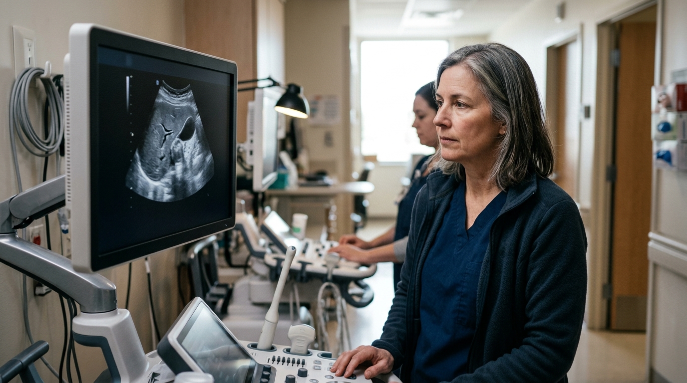
A bright liver on an abdominal scan is one of the most common incidental imaging findings, and the wording of your report matters more than the phrase itself.
If your ultrasound report says "fatty liver," "hepatic steatosis" or "increased echogenicity," it means the sonographer saw liver tissue that looks brighter than expected because fat has built up inside liver cells. It is a description of how the liver looks on the scan, not a diagnosis on its own, and it does not tell you how much fat there is or whether the liver is inflamed or scarred.
What happens next usually depends on your blood tests, your other health conditions and the specific recommendation your radiologist wrote at the end of the report.
What This Finding Is
Sound waves bounce off fat-filled liver cells differently than off normal ones. On abdominal ultrasound images, steatotic (fatty) livers look brighter than normal livers, while cirrhotic livers look lumpy and shrunken (Radiological Society of North America and American College of Radiology, 2024-05-30). The finding involves the liver tissue itself — the hepatocytes — not a lump, mass or growth.
Some fat in liver cells is typical; too much can be a problem (National Library of Medicine, MedlinePlus, 2026-07-10). Radiology sources define fatty liver disease as normal liver tissue replaced by more than 5–6% fat (Radiological Society of North America and American College of Radiology, 2024-05-30). Ultrasound cannot measure that percentage; it can only say the liver looks brighter than expected, and different reports and different machines may describe the same liver in slightly different words.
As a general pattern rather than a prediction about you: the estimated global prevalence of fatty liver disease among adults is 32%, higher among males (40%) than females (26%) (Clinical and Molecular Hepatology, 2022-12). Simple fatty liver — fat with little or no inflammation or damage — usually does not get bad enough to cause liver damage or complications (National Library of Medicine, MedlinePlus, 2026-07-10). A single ultrasound finding is a starting point for questions, not an answer.
Why This Is Sometimes Found
Common explanations are listed first; less common ones follow and are labelled as such.
- Metabolic risk factors. Fat in the liver is more common in people with type 2 diabetes or prediabetes, obesity, high cholesterol or triglycerides, high blood pressure, metabolic syndrome, or who are middle aged or older (National Library of Medicine, MedlinePlus, 2026-07-10). This pattern is called metabolic dysfunction-associated steatotic liver disease (MASLD) — the term that replaced NAFLD (American Association for the Study of Liver Diseases).
- Alcohol use. Alcohol-associated liver disease (ALD) is the other main type of steatotic liver disease (National Library of Medicine, MedlinePlus, 2026-07-10). Reports often cannot tell the two apart by appearance alone.
- Rapid weight loss, which is listed among the situations associated with liver fat (National Library of Medicine, MedlinePlus, 2026-07-10).
- Certain medicines, including corticosteroids and some cancer medicines (National Library of Medicine, MedlinePlus, 2026-07-10). Do not change anything you take — bring the list to your doctor instead.
- Less common: inflammation and scarring (MASH). MASH may involve inflammation and liver damage in addition to fat, which can lead to fibrosis and sometimes cirrhosis or liver cancer (National Library of Medicine, MedlinePlus, 2026-07-10). Ultrasound brightness alone cannot show whether this is present.
- Less common: other causes, such as certain infections including hepatitis C, or exposure to certain harmful substances (National Library of Medicine, MedlinePlus, 2026-07-10).
Size, Features, and What They Suggest
| Feature or size range (mm or cm) | What it often suggests | Typical follow-up |
|---|
| Mildly increased echogenicity, no liver enlargement reported | A small amount of liver fat; often incidental | Blood tests and risk-factor review with the ordering doctor |
| Moderate or marked brightness, with deep structures poorly seen | More fat obscuring the ultrasound beam; degree still estimated, not measured | Blood tests, often liver stiffness testing |
| Liver measured as enlarged in cm on the report (hepatomegaly) alongside brightness | Fat accumulation with increased liver size | Discussed with the ordering doctor; sometimes a specialist referral |
| Liver fat above 5–6% on MRI | Considered abnormal by radiology sources (RSNA and ACR, 2024-05-30); MRI is the most sensitive test for steatosis and can calculate a fat percentage | Used to confirm and quantify what ultrasound suggested |
| Lumpy, nodular or shrunken liver contour | Can indicate advanced fibrosis or cirrhosis (RSNA and ACR, 2024-05-30) — less common than simple brightness | Usually specialist (hepatology or gastroenterology) review |
| Any feature, any size | Follow-up timing depends on the radiologist's specific recommendation written in your own report | Follow the interval and test named in your report, and ask your doctor to explain it |
Thresholds such as 5–6% are typical cut-offs; your own report or lab may use a different range, because ranges vary by method, scanner and institution.
Frequently asked questions
Does "fatty liver" on an ultrasound mean I have liver disease?
Not by itself. The report describes how the tissue looks; a diagnosis needs your history, blood tests, exclusion of other causes and sometimes further imaging (American Association for the Study of Liver Diseases). Say that the finding can indicate excess liver fat, not that it proves a condition.
Why do I feel fine if my liver looks abnormal?
Both MASLD and alcohol-associated liver disease usually have few or no symptoms, and when symptoms occur they may be tiredness or discomfort in the upper right abdomen (National Library of Medicine, MedlinePlus, 2026-07-10). Many people with liver fat or fibrosis do not realise it because symptoms are vague (RSNA and ACR, 2024-05-30). Feeling well does not mean the finding should be ignored, and it does not mean it is harmless.
What symptoms mean I should call before my scheduled follow-up?
Contact your doctor promptly rather than waiting if you develop yellowing of the eyes or skin, swelling of the abdomen or legs, confusion, vomiting blood or black stools, severe or worsening right upper abdominal pain, or unintended appetite and weight loss — these are among the features described as appearing with progression (RSNA and ACR, 2024-05-30). It is reasonable to wait for the booked appointment if you feel the same as before the scan and have only mild fatigue.
Will I need an MRI or a biopsy?
Often neither. MRI is the most sensitive imaging test for steatosis and can calculate the percentage of liver fat, with more than 5–6% considered abnormal (RSNA and ACR, 2024-05-30), and elastography can check stiffness without a needle. Biopsy is reserved for cases where confirming the diagnosis or assessing damage changes the plan.
What should I bring to the appointment?
Bring the full written radiology report, any recent blood test slips, and a current list of medicines and supplements. Ask for the specific wording about brightness, liver size in cm and the radiologist's recommended interval, and ask what would prompt an earlier review.